

Intraoral Camera in Campbell, CA: See What Your Dentist Sees
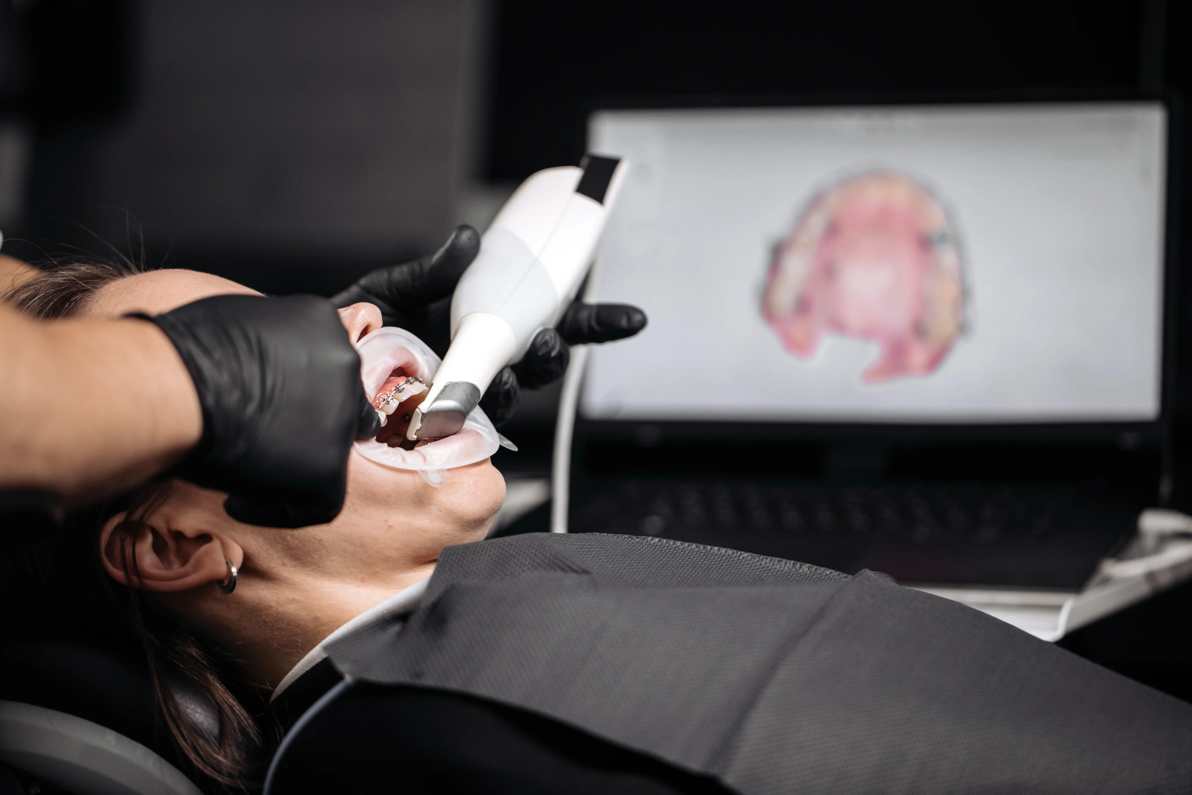
The intraoral camera gives a close-up, high-definition view of your teeth and gums. This handheld device captures magnified images in real time, helping you see what our dentist sees on a chairside screen. If you are exploring the intraoral camera in Campbell, California, this guide explains how it works, when it is used, and what those detailed images can reveal.
Intraoral Cameras Explained
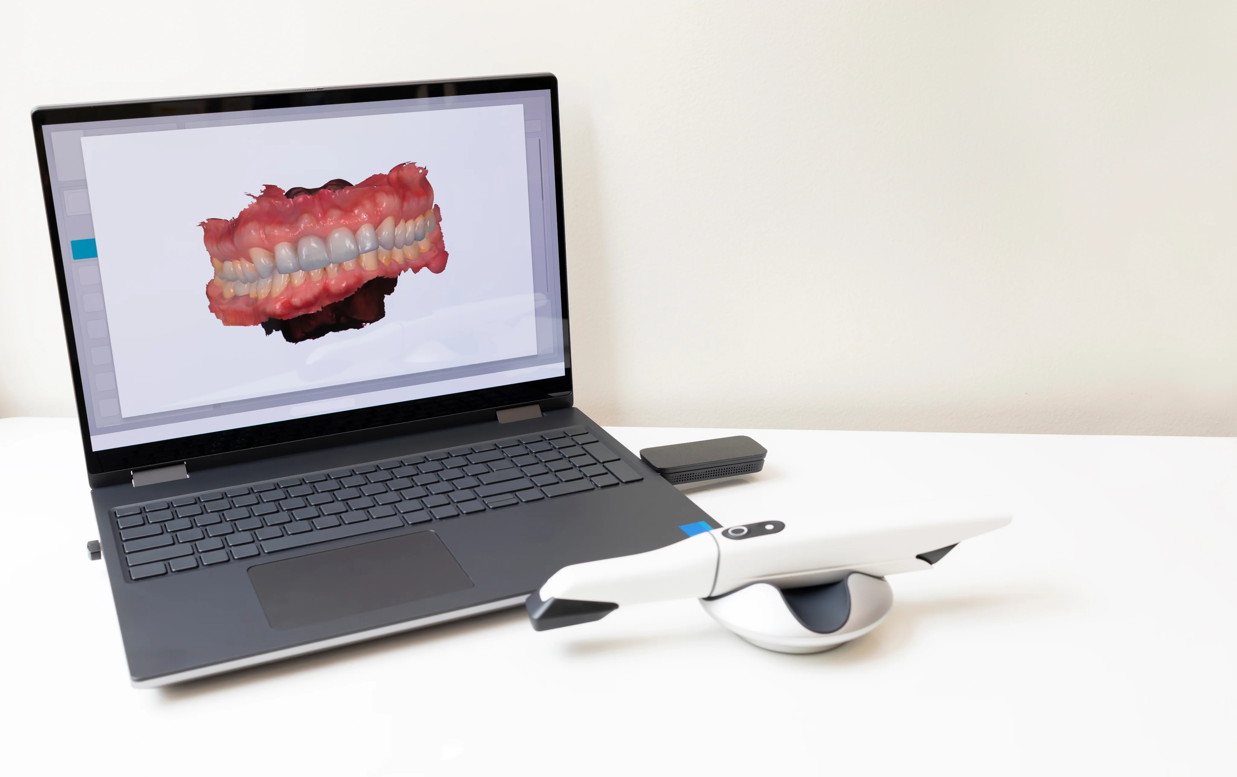
An intraoral camera is a small, wand-shaped dental imaging camera with a bright LED light and a high-quality lens. The camera takes crisp photos and short videos inside the mouth, often with magnification strong enough to highlight tiny cracks, early wear, plaque, and inflamed tissue. Images appear instantly on a monitor and can be saved to your electronic record for comparison over time. This tool is a helpful patient education device because it turns a discussion about your oral health into a visual conversation.
Unlike X-rays, an intraoral camera does not use radiation and cannot see inside teeth or bone. It excels at photographing surfaces above the gumline and at the gumline, while radiographs remain essential for viewing tooth roots, areas between teeth, and the jawbone. Together, these tools create a more complete picture of your oral health.
Benefits of Intraoral Camera Imaging
- Clearer understanding of problem areas because you can see them up close.
- Early visibility of chips, fractures, and early enamel wear before they worsen.
- Digital dental documentation that tracks changes across visits.
- Comfortable, noninvasive imaging without radiation exposure.
- More precise communication with dental specialists and dental labs when needed.
- Transparent decision making because findings are visible and easy to discuss.
How the Intraoral Camera Works
The process is quick and simple. A disposable sheath is placed over the camera for cleanliness. Our dentist or clinical team gently guides the slim wand around your teeth and gums. Images appear instantly on the chairside screen, and areas of concern can be reviewed together in real time.
The wand rests near your teeth and gums without pressing or scraping, and most images take only seconds. Most people tolerate the small camera well. Slower movements, brief breaks, and nasal breathing usually make imaging more comfortable for patients with a strong gag reflex.
Images are part of your electronic record and can be shared upon request for your personal files or for coordination with specialists.
Questions About Our Intraoral Camera?
If you have questions about intraoral cameras in Campbell, CA, or how this patient education tool fits into your exam, contact Kirkwood Dental at 408-378-8500 to schedule with Dr. Vinh T. Pham.
Frequently Asked Questions About Intraoral Camera
- Yes. It uses light and optics, not radiation. A fresh, single-use sheath is placed for each patient to support infection control.
- The camera shows color images of tooth and gum surfaces, while X-rays show internal structures like roots and bone. Dentists often use both for a complete evaluation.
- No. The process is noninvasive. The wand rests near your teeth and gums without pressing or scraping, and most images take only seconds.
- Yes. Images are part of your electronic record and can be shared upon request for your personal files or for coordination with specialists.
- It can reveal visible signs of decay, staining, or softened enamel on surfaces. However, X-rays are still important for finding hidden cavities between teeth.
- Most people tolerate the small camera well. Slower movements, brief breaks, and nasal breathing usually make imaging more comfortable.
If you have questions about intraoral cameras in Campbell, CA, or how this patient education tool fits into your exam, contact Kirkwood Dental at 408-378-8500 to schedule with Dr. Vinh T. Pham.